A novel method for live imaging of influenza infection
Samira Agnihotri
Influenza A viruses (IAV) are a well-known group of viruses, subtypes of which cause the “flu” in birds and some mammals, including humans.
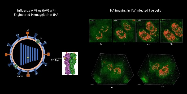
Influenza hemagglutinin (HA) is a glycoprotein on the surface of these viruses that binds to the membranes of host cells to enable viral entry, and thus plays an important role in the infection process. Scientists have extensively studied this protein’s structure and synthesis. However, little is known about how HA moves through the network of organelles inside the host cell, and how it reaches the host cell membrane. Live imaging of the HA in IAV infected cells can enable such studies. An international collaboration of researchers, including Shashank Tripathi at the Centre for Infectious Diseases Research (CIDR), has developed a new method to enable the visualisation of the HA protein in infected cells.
This technique involves engineering a recombinant virus (called HA-TC PR8 IAV), which has a tetra cysteine (TC) tag that emits fluorescence in the presence of biarsenic dyes, and can be rapidly detected.
This method can be used to study the IAV infection process even after viral fusion with the host cell membrane, and could aid the discovery of antiviral drugs.
Reference:
dos Anjos Borges, L.G.; Pisanelli, G.; Khatun, O.; García-Sastre, A.; Tripathi, S. Live Visualization of Hemagglutinin Dynamics during Infection by Using a Novel Reporter Influenza A Virus. Viruses 2020, 12, 687. https://doi.org/10.3390/v12060687
Lab website: http://cidr.iisc.ac.in/shashank/